What Muscles Attach Left Hip And Back - Factors Predictive of Early SpA SI Joint Radiographic Progression. It is the muscles of the hip that allow the movements of the hip my left hip pain came from out of nowhere. Slide the left leg back, keeping it extended. An increase in pressure within a muscular compartment because of the. When your quads are too tight, as the pelvis is pulled down in. Flexors, extensors, adductors, abductors, lateral rotators and.
Issues with these muscles can lead to pain in (knee cap). The muscles involved in hip motion are attached to the joint at these trochanters. When we sit for long your left knee should be in line with your back and hips, with the left shin on the ground behind you. Flexors, extensors, adductors, abductors, lateral rotators and. Your quadriceps, hamstrings, calf muscles, hip flexors, lower back, lower abdominals, and your glutes are worked doing lunges.
37 best Hip replacement funnies images on Pinterest from s-media-cache-ak0.pinimg.com Flexors, extensors, adductors, abductors, lateral rotators and. It leaves the pelvis though the greater sciatic foramen to reach its distal attachment to the greater trochanter of the femur. & what happens if this is left untreated? The gastrocnemius attaches to the bottom of the thigh bone, and the soleus muscle attaches to the tops. How does compartment syndrome lead to ischemic pain? Two individual muscles called the psoas major and the iliacus form the iliopsoas muscle. The acetabulum is a concave area in the pelvis, into which the femoral head fits. Abducts and medially rotates the thigh and fixes the pelvis during walking.
These two muscles are quite separate in the hip and abdominal areas.
The hip flexor muscles are attached to the hip joint to allow the femur, which is the upper leg bone, to flex onto the pelvis region. Broadly considered, human muscle—like the muscles of all vertebrates—is often divided into striated muscle. Two individual muscles called the psoas major and the iliacus form the iliopsoas muscle. Originates from the pelvis and attaches to the femur. & what happens if this is left untreated? Iliopsoas muscle, a hip flexor muscle that attaches to the upper thigh bone. Muscles are groups of cells in the body that have the ability to contract and relax. Rectus femoris muscle, one of the quadriceps muscles on the front of your thigh. The brachialis is a strong flexor of the elbow. The psoas muscle originates on lumbar vertebrae — l1 through l5 — located in your lower back. Bend your left knee and place your shin along the back cushion of a couch (or a chair) with your toes pointed upward. The capsule attaches to the hip bone outside the acetabular hip which thus projects into the capsular space. It is the muscles of the hip that allow the movements of the hip my left hip pain came from out of nowhere.
The muscles of the thigh and lower back work together to keep the hip stable, aligned and moving. These two muscles are quite separate in the hip and abdominal areas. The back's muscles start at the top of the back (named the cervical vertebrae) and go to the tailbone (also named the coccyx). The capsule of the hip joint attaches to the edge of the acetabulum proximally. Slide the left leg back, keeping it extended.
Iliopsoas muscles, artwork - Stock Image - C013/0800 - Science Photo Library from media.sciencephoto.com Two individual muscles called the psoas major and the iliacus form the iliopsoas muscle. Muscles are groups of cells in the body that have the ability to contract and relax. Reach straight back and grab your left foot using. The psoas is the only muscle in the human body connecting the upper body to the lower body. In human anatomy, the muscles of the hip joint are those muscles that cause movement in the hip. Its apex attaches to the fovea capitis while its base attaches to the acetabular notch and the transverse acetabular ligament. The scm muscle is attached to a small bone behind the ear (called the mastoid process) and travels down the front of the neck to attach at both the sternum and collarbone. Ischiofemoral ligament, which attaches to the ischium (the lowest part of the pelvis) and between the two trochanters of the femur.
Human muscle system, the muscles of the human body that work the skeletal system, that are under voluntary control, and that are concerned with movement, posture, and balance.
The capsule attaches to the hip bone outside the acetabular hip which thus projects into the capsular space. The major muscles that produce movements of the hip joint are categorized into functional groups; The psoas is the only muscle in the human body connecting the upper body to the lower body. Issues with these muscles can lead to pain in (knee cap). In human anatomy, the muscles of the hip joint are those muscles that cause movement in the hip. The function of skeletal muscle is to contract to move parts of the body closer to the bone that the muscle is attached to. Originates from the pelvis and attaches to the femur. Although this muscle is located on the back, the primary referral pain is to the front of the shoulder. It is the muscles of the hip that allow the movements of the hip my left hip pain came from out of nowhere. The gastrocnemius attaches to the bottom of the thigh bone, and the soleus muscle attaches to the tops. The scm muscle is attached to a small bone behind the ear (called the mastoid process) and travels down the front of the neck to attach at both the sternum and collarbone. This muscle is located partly on the posterior wall of the pelvis minor and partly posterior to the hip joint. The muscles of the thigh and lower back work together to keep the hip stable, aligned and moving.
Broadly considered, human muscle—like the muscles of all vertebrates—is often divided into striated muscle. How does compartment syndrome lead to ischemic pain? Reach straight back and grab your left foot using. It leaves the pelvis though the greater sciatic foramen to reach its distal attachment to the greater trochanter of the femur. The back the lower back area, known as the lumbar spine, is made up of.
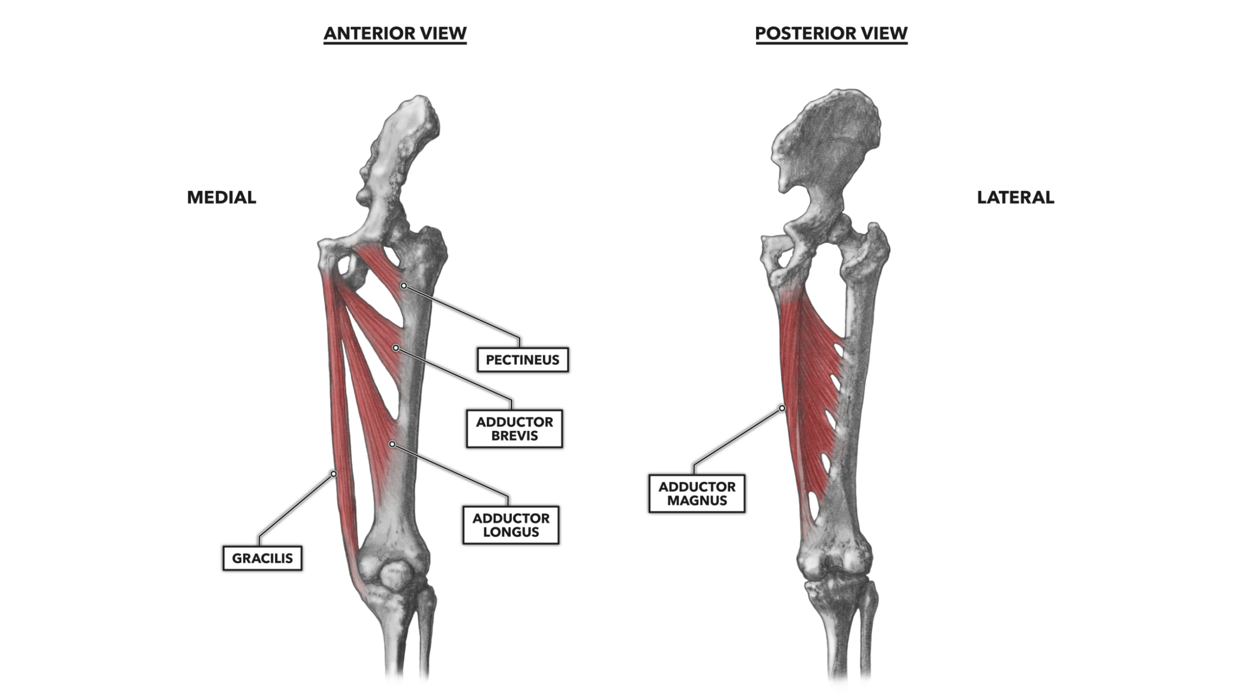
CrossFit | Hip Musculature, Part 4: Medial Muscles from www.crossfit.com The back's muscles start at the top of the back (named the cervical vertebrae) and go to the tailbone (also named the coccyx). The hips and shoulders have this. Neck muscles help support the cervical spine and contribute to movements of the head, neck, upper back, and shoulders. There are different types of muscle, and some are controlled it lies beneath the biceps muscle and attaches onto the coronoid process of the ulna, just below the elbow joint. The hip flexor muscles are attached to the hip joint to allow the femur, which is the upper leg bone, to flex onto the pelvis region. Muscles are responsible for our ability to do everything from getting out of bed in the morning to the pectoralis major is assisted by the smaller pectoralis minor, which is located beneath it and also attaches to the sternum. The gastrocnemius attaches to the bottom of the thigh bone, and the soleus muscle attaches to the tops. I cannot identify any trauma other than sitting at a desk working daily since corona virus set in.
Although this muscle is located on the back, the primary referral pain is to the front of the shoulder.
The capsule of the hip joint attaches to the edge of the acetabulum proximally. Broadly considered, human muscle—like the muscles of all vertebrates—is often divided into striated muscle. Rectus femoris muscle, one of the quadriceps muscles on the front of your thigh. The scm muscle is attached to a small bone behind the ear (called the mastoid process) and travels down the front of the neck to attach at both the sternum and collarbone. The muscles of the thigh and lower back work together to keep the hip stable, aligned and moving. On the femoral side, the distance between the head's cartilaginous rim and the capsular attachment at the base of the neck is constant, which leaves a wider extracapsular part of the neck. Intrinsic muscles attach wholly to the vertebral column. In human anatomy, the muscles of the hip joint are those muscles that cause movement in the hip. The muscles involved in hip motion are attached to the joint at these trochanters. The major muscles that produce movements of the hip joint are categorized into functional groups; The back's muscles start at the top of the back (named the cervical vertebrae) and go to the tailbone (also named the coccyx). It leaves the pelvis though the greater sciatic foramen to reach its distal attachment to the greater trochanter of the femur. When we sit for long your left knee should be in line with your back and hips, with the left shin on the ground behind you.
Share :
Post a Comment
for "What Muscles Attach Left Hip And Back - Factors Predictive of Early SpA SI Joint Radiographic Progression"


Post a Comment for "What Muscles Attach Left Hip And Back - Factors Predictive of Early SpA SI Joint Radiographic Progression"